
Anatomy of a Neuron
Explore the structure of a multipolar neuron, including dendrites, axon, myelin sheath, and synapse. Diagrams cover neural impulse conduction and the stages of signal transmission.

Microscopic views and diagrams of cellular components, neurons, and brain structures — free resources for students and educators.
Browse our collection of free diagrams and articles covering the microscopic world of cells. From the intricate structure of neurons to the functional regions of the brain and the pathways of blood vessels, these resources support study and teaching in cell biology.
Cell anatomy opens a window into the smallest living units that build every tissue and organ in the human body. Beneath a microscope, these structures reveal membranes, organelles, and internal pathways that coordinate growth, repair, and communication. Educational diagrams simplify these complex arrangements into clear visual references, helping learners connect abstract biological concepts with the shapes and patterns they observe in laboratory settings. Such resources support classroom teaching and independent study alike, offering a steady foundation for understanding how cellular form relates to function in health, development, and disease across plants, animals, and humans.
Neurons stand out among specialized cells because their extended branches allow rapid signaling across the nervous system. A multipolar neuron typically shows a central cell body surrounded by branching dendrites, a long projecting axon, and an insulating myelin sheath that supports swift impulse transmission. Synaptic terminals at the axon tip pass signals to neighboring cells, completing networks that govern sensation, thought, and movement. Diagrams of these features help students visualize how structure supports electrical communication, turning the nervous system into a topic that feels tangible and approachable rather than abstract.
Brain anatomy broadens the cellular view into organized regions responsible for memory, language, coordination, and emotion. Cortical layers, deep nuclei, and connecting pathways illustrate how groups of neurons cooperate to produce complex behavior. Comparative studies between species also reveal how brain architecture adapts to different sensory demands and environmental pressures, enriching lessons in physiology and evolution. Visual resources linking microscopic cell features to larger brain regions help learners appreciate the layered design of the nervous system, from individual synapses to integrated circuits that shape perception and conscious experience.
Blood vessels form another essential study area, weaving through tissues to deliver oxygen, nutrients, and signaling molecules while removing metabolic waste. Diagrams often trace branching patterns from major arteries down to fine capillary networks, showing how cellular respiration depends on this circulatory reach. Together with neuron and brain illustrations, vascular imagery completes a more holistic picture of human anatomy, encouraging students to see the body as a coordinated system where microscopic structure, regional organization, and physiological function work in continuous harmony.
Explore the structure of a multipolar neuron, including dendrites, axon, myelin sheath, and synapse. Diagrams cover neural impulse conduction and the stages of signal transmission.
Labeled diagrams of brain structures and their physiological roles. Includes brain anatomy quizzes, MRI views, and functional region maps for study and review.
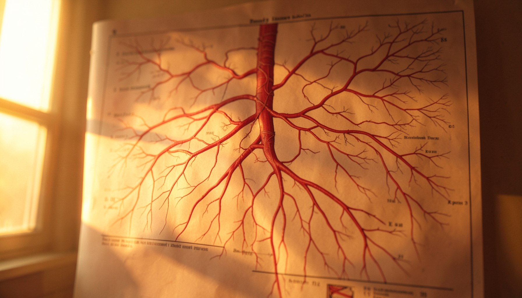
Review sheets and exercises covering blood vessel anatomy, including heart vessels, blood flow tracing, and vessel identification. Suitable for self-assessment and classroom use.